Diffraction pattern from a double helix
Friedrich Zemlin
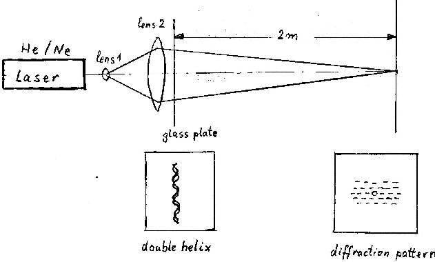
The following description is an experiment on a model carried out with a laser beam. First an appropriate double helix was manufactured by turning two Nylon strings. The Nylon strings, as used for fishing, have a diameter of 0.2mm.This double helix was then used for Laser diffraction as seen in the sketch of the laser optical bench (Fig.1).
Figure 1
The resulting diffraction pattern from this double helix is shown in Fig 2.
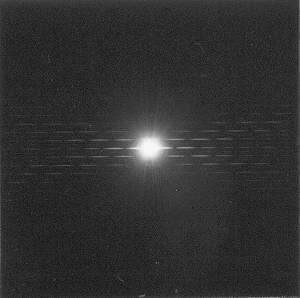
Figure 2
To distinguish between right handed turned and left handed turned double helices, we immersed in further experiments half of the double helix string into a film of oil, which was brought onto the supporting glass plate. The diffraction patterns are shown in Fig.3 and Fig.4 .
![]()
![]()
Figure 3 (right handed) Figure 4 (left handed)
This experiment was done many years ago as a preliminary study, we were really interested in electron optical imaging and diffraction of DNA crystals at very low temperatures using a Helium cooled cryo-electronmicroscope. But we gave up these experiments, because we had the feeling, even if such experiments would be successful no thrilling news would come out. May be these experiments can be a help for students to understand the diffraction pattern of double helices.
![]()